
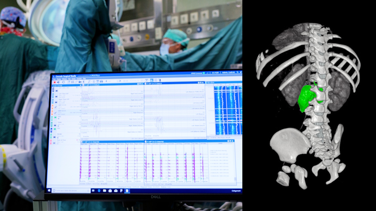
A little girl of just over a year old was subjected to the Baby Jesus of Rome to a delicate intervention for the complete removal of a neuroblastomaa form of tumor which in his case had reached the spinal canal and compressed a nerve involved in the movement of a leg. The operation, which lasted about four hours, was prepared through three-dimensional reconstructions and virtual simulations of the intervention.
Neuroblastoma near the nerves, surgery is simulated in 3D at Baby Jesus before surgery
The particular conformation of the mass required modifying the normal surgical approach. Rather than reaching the tumor through the abdomen, the team chose an approach from the back, considered more suitable to operate simultaneously on the external part of the mass and on the portion penetrated into the spinal canal. A choice made possible by the preliminary study of the patient’s anatomy through three-dimensional models.
Before the operation, the doctors virtually reconstructed the relationships between the tumor, the spine, the spinal cord and the nervous structures. The simulations allowed us to evaluate different possibilities and identify the most effective surgical route to reach the mass while reducing the risk of damaging the nerves.
«The location of the tumor forced us to completely rethink the surgical approach. The three-dimensional simulations allowed us to study each phase of the operation in advance and to choose an access from the back, unusual for this type of tumor but the most suitable in this case. The goal was to completely remove the disease while preserving the most important nervous structures.”», he explained Giorgio Persanoof the Bambino Gesù Oncology Surgery Simple Operational Unit. The objective was to completely remove the disease while preserving the most important nervous structures.
The procedure required the collaboration of various skills. Oncology surgeons, neurosurgeons, neurophysiologists and anesthetists worked together during the different phases of the operation. A central role was entrusted to intraoperative neurophysiological monitoring, which allowed the activity of nervous structures to be monitored in real time while surgeons operated in proximity to the nerves.
From 3D simulation to the operating room: this is how technology helped remove a complex neuroblastoma
The management of anesthesia was also calibrated to the needs of the operation. The child underwent total intravenous anesthesia with dosages and infusions adjusted to maintain clinical stability and, at the same time, preserve the reliability of the signals used by neurophysiological monitoring. The dialogue between the anesthesiologist and the neurophysiologist was therefore constant for the entire duration of the procedure.
«When a tumor grows so close to the nerves that control the movement of a leg, every millimeter counts. Neurophysiological monitoring allows us to verify in real time that these structures continue to function while the surgeon works. By integrating neurosurgery, neurophysiology, anesthesia and oncological surgery we can tackle extremely delicate interventions with a higher level of safety”said Dr Andrea Carairesponsible for the complex operational unit of
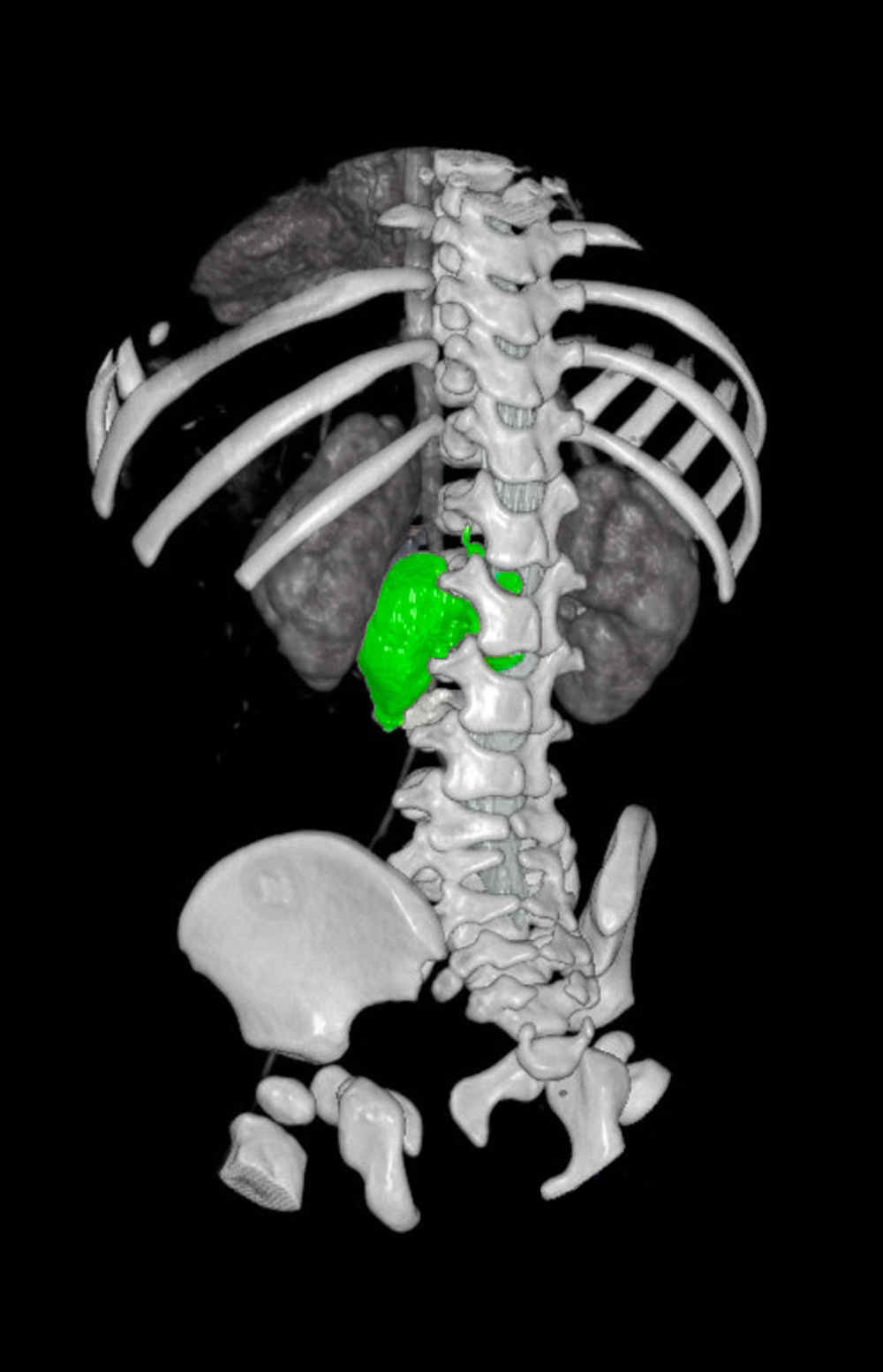
3D neuroblastoma
Neuroblastoma is a malignant tumor that originates from cells of the sympathetic nervous system and represents the most common extracranial solid tumor in children under five years of age. It can develop in different areas of the body, including the adrenal glands and areas along the spine.
In the case of the little patient, a diagnostic biopsy and the chemotherapy foreseen by the protocols were carried out before the operation. The reduction of the mass obtained with the treatment allowed us to proceed with the planning of the surgical intervention. For more information, find the Baby Jesus Press Release here with supporting videos and images.
The removal was completed successfully. Six days after the operation the little girl was discharged from hospital and began the rehabilitation process. A result that shows how the combination of digital planning, specialist surgery and continuous monitoring could also allow us to tackle pediatric tumors located in particularly delicate areas, where a few millimeters can make the difference.